~A Boy Always Young~

2015年3月10日 星期二

Charcot’s Neuroarthropathy


Charcot’s neuroarthropathy affects the joints and bones of the feet. The development of this condition include severe peripheral neuropathy and autonomic dysfunction with increased blood flow to the foot; the peripheral circulation is usually intact.

The actual pathogenesis is poorly understood; however, the patient is vulnerable to trauma that may not recall. Repetitive trauma results in increased blood flow through the bone, increased osteoclastic activity, and remodeling of bone. In certain cases, patients walk on a fracture, which leads to continuing destruction of bones and joints in that area. Acute Charcot’s neuropathy may be triggered by any event that leads to localized inflammation. This may trigger a vicious cycle in which there is increasing inflammation, increasing expression of RANKL (a member of the tumor necrosis factor superfamily), and increasing bone breakdown.

Charcot’s neuropathy is sometimes difficult to distinguish from osteomyelitis or an inflammatory arthropathy. A unilateral swollen, hot foot in a patient with neuropathy must be considered to be a Charcot foot.

Charcot’s arthropathy can be diagnosed in most patients by plain radiography and a high index of suspicion. Radiographs may reveal bone and joint destruction, fragmentation, and remodeling. In such cases, the three-phase bisphosphonate bone scan shows increased bone uptake, although the 111In-labeled bone scan will be negative in the absence of infection.

Management of the acute phase involves immobilization. Evidence suggests that treatment with bisphosphonates, which reduce osteoclastic activity, may reduce swelling, discomfort, and bone turnover markers. Although Charcot’s neuroarthropathy is rare, it should be suspected in any patient with unexplained swelling and heat in a neuropathic foot.
read more

Glucose clamp technique


Glucose clamp technique is used to measure either how well an individual metabolizes glucose or how sensitive an individual is to insulin. Two types of clamps are quite commonly used.

The hyperglycemic clamp, which requires maintaining a high blood sugar level by perfusion or infusion with glucose, is a way to quantify how fast beta-cells respond to glucose.

The hyperinsulinemic clamp, which requires maintaining a high insulin level by perfusion or infusion with insulin, is a way to quantify how sensitive the tissue is to insulin.

Hyperglycemic clamp technique:
Glu raised to 125 mg/dl above basal levels by a continuous infusion of glucose. The glucose infusion rate is an index of insulin secretion and glucose metabolism. The hyperglycemic clamps are often used to assess insulin secretion capacity.

Hyperinsulinemic-euglycemic clamp technique:
The plasma insulin concentration is acutely raised and maintained at 100 μU/ml by a continuous infusion of insulin. The plasma glucose concentration is held constant at basal levels by a variable glucose infusion. When the steady-state is achieved, the glucose infusion rate equals glucose uptake by all the tissues in the body and is therefore a measure of tissue insulin sensitivity. The hyperinsulinemic clamps are often used to measure insulin resistance.
read more

Struma Ovarii


A teratoma of the ovary contains thyroid tissue that becomes hyperactive. Mild features of thyrotoxicosis result, such as weight loss and tachycardia, but there is no evidence of goiter or eye signs. Serum FT4 and T3 are mildly elevated, serum TSH is suppressed, and RAIU over the neck is low. Total body scan reveals uptake of radioiodine in the pelvis, rather than in the neck. The disease is curable by removal of the teratoma.
read more

2015年3月9日 星期一

MIBG scan


整理自長庚資料 (http://kelwww.cgmh-mi.com/examnm/i-131-mibg-scan)

MIBG(Metaiodo Benzylguanidine),是一種類似正腎上腺素的藥劑,會聚積在儲有catecholamine的腎上腺髓質嗜鉻性細胞或是交感神經末梢內,診斷嗜鉻性細胞瘤有76~100%的敏感度,在臨床上最常用於檢查的便是嗜鉻性細胞瘤。為預防I-131聚積而傷害甲狀腺,藥物注射前三天得開始服用 Lugol's solution(含碘試劑,用來阻斷解離的放射碘傷害正常甲狀腺組織)。MIBG因為結構類似 norepinephrine,必須緩慢注射以免引起高血壓的危險性。

適應症
嗜鉻性細胞瘤(pheochromocytoma)
甲狀腺髓質癌(medullary thyroid carcinoma)
亞神經節瘤(paragangliomas)
神經母細胞瘤(neuroblastoma)
類癌瘤(carcinoid tumor):位於小腸、闌尾、胃結腸發生之一種界線分明的黃色瘤。
read more

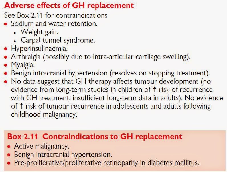
Adverse effects of GH replacement


read more

Pendred syndrome


Pendred syndrome is characterized by both goiter and sensorineural deafness (not caused by hypothyroidism). It is caused by a biallelic mutation in the SLC26A4 gene, located on chromosome 7 (7q31). Pendrin, a multifunctional anion exchanger, functions in both the thyroid gland and inner ear.

In the apical membrane of the thyroid gland, pendrin facilitates transport of iodide into the exocytic vesicles in which thyroid hormone is synthesized.

Pendrin is also expressed in the inner ear and is important for normal endolymph composition and maintenance of the endocochlear potential.
read more

MELAS


MELAS=The syndrome of mitochondrial encephalomyopathy with lactic acidosis and stroke-like episodes

An A-to-G transition in the mitochondrial transfer RNA Leu(UUR) gene at base pair 3243 has been shown to be associated with maternally transmitted diabetes and sensorineural hearing loss. In other subjects, this mutation is associated with diabetes and the syndrome of mitochondrial myopathy, encephalopathy, lactic acidosis, and stroke-like episodes (MELAS syndrome). The mitochondrion plays a key role in the regulation of insulin secretion, particularly in response to glucose.

MELAS is a maternally inherited multisystemic disorder caused by mutations of mitochondrial DNA. The hallmark is the occurrence of stroke-like episodes that result in hemiparesis, hemianopia, or cortical blindness. Other common features include focal or generalized seizures, recurrent migraine-like headaches, vomiting, short stature, hearing loss, and muscle weakness. 80 percent of cases are related to the A3243G mutation and 10 percent to the T3271C transfer RNA mutation.

MELAS usually manifests in childhood after a normal early development [46,47,50]. A relapsing-remitting course is most common, with stroke-like episodes leading to progressive neurologic dysfunction and dementia.

The diagnostic criteria required:
--stroke-like episodes before age 40 years,
--encephalopathy characterized by seizures or dementia,
--either blood lactic acidosis or the presence of ragged red fibers in skeletal muscle biopsy

However, a broader range of phenotypes is now recognized as compatible with the diagnosis, including clinical onset after age 40.
read more

Other Causes of Hypoparathyroidism


Neonatal hypoparathyroidism can be part of the DiGeorge syndrome (dysmorphic facies, cardiac defects, immune deficiency, and hypoparathyroidism) due to a microdeletion (or other genetic abnormalities) on chromosome 22q11.2.

The HDR syndrome (hypoparathyroidism, sensorineural deafness, and renal anomalies) due to mutations in or deletion of an allele of the GATA3 transcription factor; and other rare conditions.
read more

2015年3月5日 星期四

Primary aldosteronism


Oral sodiumloading test
病人接受連續三天共218 mmol之鈉鹽(約合12.8公克氯化鈉)之高鈉飲食18
,在第三天到第四天間收集二十四小時小便測量尿液中的醛固酮、鈉離子及肌酸酐(creatinine)。為確認體內鈉鹽是足夠的,小便中之鈉離子應超過每天200 mmol,而尿液醛固酮超過每天33.3 nmol(約合12 μg)即可診斷

Intravenous saline infusion test
overnight空腹的情況下,兩公升生理食鹽水以超過四小時之流速靜脈輸注於側躺之病人,完成輸注時,抽血檢查病人之血清醛固酮濃度;若血清醛固酮濃度超過10 ng/dL,則可診斷為原發性高醛固酮症;介於5~10 ng/dL則可能為雙側腎上腺增生;而正常人小於5 ng/dL

Fludrocortisone suppression test
Fludrocortisone acetate每六小時0.1mg共四天,同時照三餐每餐給予氯化鈉鹽片2公克。在第四天抽血檢查病人之血清醛固酮濃度,若高於6 ng/dL則可診斷原發性高醛固酮症。

Captopril test
在採檢血清醛固酮濃度及血清張力素活性前一 ~ 二小時單次口服captopril 25 ~ 50 mg,在正常情形下血清醛固酮濃度受captopril抑制會下降超過30%;但原發性高醛固酮症患者則否,且其血清張力素活性仍持續受到抑制。

Posture change test

病人前一晚維持臥姿睡眠,隔天抽晨血後,持續站姿2~4小時再抽第二次血,比較兩次之血清醛固酮濃度,若第二次高於第一次50%以上則為陽性反應;大部分雙側腎上腺增生呈陽性反應,但醛固酮分泌腺瘤及醣皮質醇可矯正型皮質醛酮症其第二次血清醛固酮濃度不會上升,反而下降或不變,故姿勢變化檢查為陰性。
read more